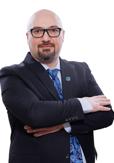
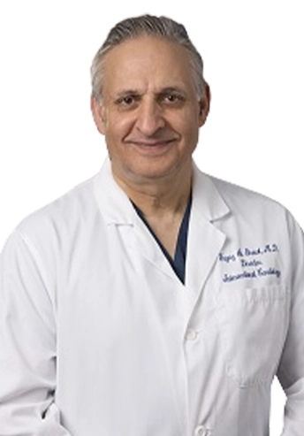
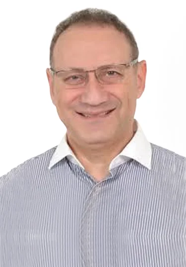
The Cardiac Center Of Excellence
Zulekha Hospital has been at the heart of providing the finest cardiac care in UAE since 2007 with a team of dedicated international experts together with advanced technology and world class facilities.
The department is fully equipped for non-invasive and invasive investigations such as Transthoracic and Transesophageal echocardiography and treadmill testing. The Cardiac Center of Excellence boasts of a top-notch digital cardiac catheterization lab and has been routinely performing diagnostic and interventional procedures. Alongside the routine diagnostic invasive procedures like angiography and catheterization studies, the center performs coronary and peripheral angioplasty (angioplasties of the leg for other blood vessels), with stenting and balloon valvuloplasty for all valvular lesions. Device and coil closure of PDA and device closure of ASD are being performed on patients of all ages.
The range of specialized care for screening, diagnosis and treatment of various heart conditions are as follows:
Non Invasive Cardiology
- 24 hour Holter monitor
- 2D Echocardiogram
- Transesophageal Echocardiogram
- Exercise / Dobutamine stress Echocardiogram
- Exercise Stress Test
Invasive Cardiology
- Cardiac catheterization
- Coronary intervention
- Peripheral angiogram and interventions: carotid, renal, celiac, mesenteric and extremities
- Permanent pacemaker placement
- AICD / CRT placement
- ASD/ PFO closure
Additionally, the Cardiac Center of Excellence at Zulekha Hospital Sharjah is fully equipped to facilitate high-risk treatments locally including procedures such as Cardiothoracic and Vascular Surgery (CTVS) and Coronary Artery Bypass Grafting (CABG) to improve blood flow to the heart.
Our surgeons will provide complete evaluation, diagnosis, and surgical treatment for a wide variety of disorders. Our goal will be to provide contemporary, evidence-based Cardiothoracic and Vascular surgery in a personalized fashion. Using an integrated approach in the treatment of heart, lung and Vascular disease, our surgeons will work closely with cardiologists, pulmonologists, endocrinologists and other specialists to make one’s health our top priority.
Procedures that will be performed:
Adult Cardiac Surgery
Coronary Artery Bypass Surgery
Bypass surgery is performed frequently nowadays, both in men and women. Bypass surgery is meant for patients with clogged (coronary) arteries of the heart, where medications, ballooning or stenting cannot help anymore. Bypass surgery in advanced cases of heart artery disease can bethemore promising solution to pursue. Bypass surgery in our times can be performed safely and efficiently, at very low risk, depending on every patient's history and concurrent problems.
Reason for the Procedure
- One of the most common and effective procedures to manage blockage of blood to the heart muscle.
- Improves the supply of blood and oxygen to the heart.
- Relieves chest pain (angina).
- Reduces risk of heart attack.
- Improves ability for physical activity that has been limited by angina or ischemia
Valvular Heart surgery
Heart valve surgery is a procedure to treat heart valve disease. Heart valve disease involves at least one of the four heart valves not working properly. Heart valves keep blood flowing in the correct direction through your heart.
The four valves are the mitral valve, tricuspid valve, pulmonary valve and aortic valve. Each valve has flaps - called leaflets for the mitral and tricuspid valves and cusps for the aortic and pulmonary valves. These flaps open and close once during each heartbeat. Valves that don't open or close properly disrupt blood flow through your heart to your body.
In heart valve surgery, your surgeon repairs or replaces the affected heart valves. Many surgical approaches can be used to repair or replace heart valves, including open-heart surgery or minimally invasive heart surgery.
Your treatment depends on several factors, including your age, your health, the condition of the affected heart valve and the severity of your condition.
Aortic root surgery
Aortic root surgery is a procedure to treat an enlarged section, or aneurysm of the aorta. The aorta is the large blood vessel that carries blood from the heart to the body. The aortic root is located near where the aorta and heart connects.
Why it's done
Doctors perform aortic root surgery to prevent a burst aneurysm. They also perform it to prevent a tear in the inner layer of the aorta's wall (aortic dissection). They also do it to prevent the enlarged aorta from stretching the attached aortic valve. Aortic aneurysms near the aortic root may be related to Marfan syndrome and other heart or inherited conditions.
Minimally invasive heart surgery
Minimally invasive heart surgery involves making small incisions in the right side of your chest to reach the heart between the ribs, rather than cutting through the breastbone, as is done in open-heart surgery.
Minimally invasive heart surgery can be performed to treat a variety of heart conditions. Compared with open-heart surgery, this type of surgery might mean less pain and a quicker recovery for many people
Thoracic surgery
- Lung surgeries
- Surgery for lung cancer
- Video assisted Thoracic surgery
- Intercostal drainage
- Chemo port insertion for Chemotherapy
- Surgery for tuberculosis
- Decortication for thickened pleura
Vascular surgery
- Peripheral arterial bypass
- Endoscopic Vascular surgery
- Arteriovenous fistula creation for dialysis
- Treatment for Deep Venous thrombosis
- Laser therapy (EVLT) for varicose veins
- Aortic surgeries
- Treatment of Ischemic and Diabetic wounds
Paediatric Cardiology
Pediatric Cardiology is a subspecialty of pediatrics that focuses on the diagnosis and treatment of heart conditions in children, including congenital heart defects. Our pediatric cardiology department is furnished with the latest state-of-the-art technologies to diagnose and treat any type of cardiac problem in our pediatric patients. The presence of all other specialties under one roof in Zulekha Hospital ensures we can easily coordinate with children’s pediatricians and treating doctors.
Common heart problems treated in children:
- Chest pain
- Newborn screening
- Evaluation of murmur
- Fainting episodes
- Palpitations and arrythmias
- Heart failure
- Congenital heart diseases
- Pulmonary hypertension
- Cardiomyopathy
- Myocarditis
The department is equipped with the following:
- Pediatric echocardiography
- 24 hour Holter study
- Electrocardiography
- Treadmill test
Department Doctors
Dr. Chidanand Bedjirgi
Specialist Cardiothoracic & Vascular Surgeon
(Appointment is subjected to the availability of doctor)

